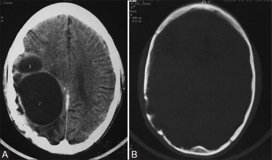
Figure 15 (A and B).

Recurrent hydatid cyst of brain. Contrast-enhanced CT images show (A) multiloculated hypodense mass with attenuation similar to cerebrospinal fluid in the right parietal lobe; minimal perilesional edema is also seen and (B) erosion of inner table of skull
