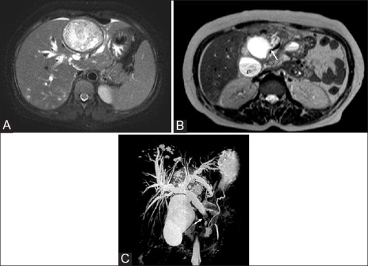
Figure 3 (A-C).

Hepatic hydatid cyst with biliary communication in a middle-aged female who presented with obstructive jaundice. (A) Axial T2W image shows a type II cyst seen as heterogeneous hyperintense mass with few daughter cysts in the left lobe of the liver. An intact dark rim of pericyst is seen. Intrahepatic biliary dilatation and small developing cysts in the posterior part of the right lobe are also seen (B) Axial T2W image through the inferior part of liver reveals cystobiliary communication (arrow), irregularity of cyst outline, and loss of dark rim (C) Coronal thick slab MR cholangiopancreatogram shows dilated intahepatic and extrahepatic bile ducts with hypointense filling defect (arrow) due to hydatid material in the distal common bile duct. Gb = Gall bladder
