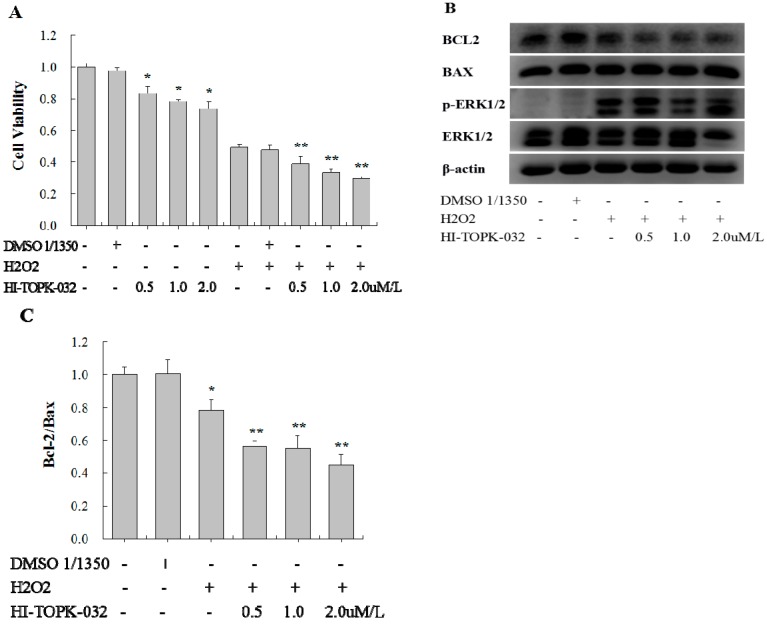
Figure 4.
Analysis of the effects of the TOPK specific inhibitor HI-TOPK-032 on cell viability, apoptosis, and protein expression. H9C2 cardiomyocytes were incubated with HI-TOPK-032 at concentrations of 0.5, 1.0, and 2.0 µM for 24 h and then stimulated with 750 µM of H2O2 for 1 h. Dimethyl sulfoxide (DMSO) was used as a solvent control. (A) Cell viability as detected by MTS assay; (B) Western blot analysis of Bcl-2, Bax, ERK, and p-ERK expression. β-actin was used as a loading control; (C) The Bcl-2/Bax ratio was calculated and compared. * p < 0.05 compared with nonstimulated controls; ** p < 0.05 compared with H2O2-stimulated controls.

