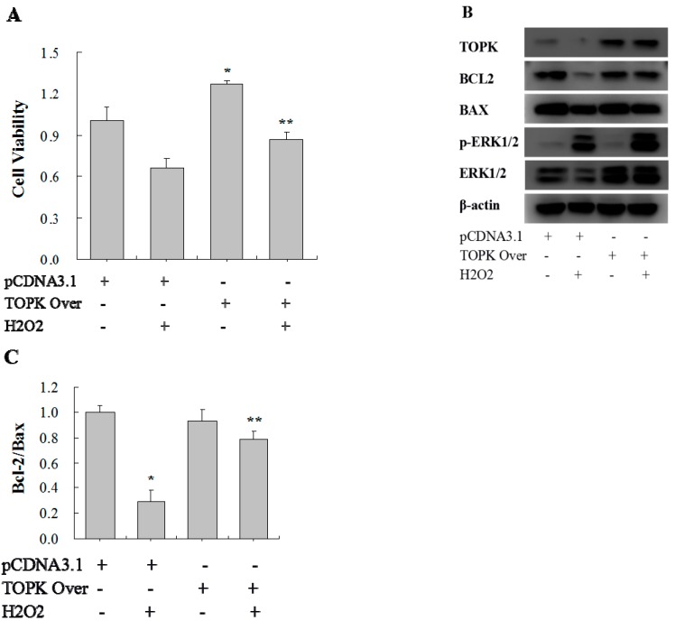
Figure 5.
Analysis of the effects of TOPK overexpression on cell viability, apoptosis, and protein expression. H9C2 cardiomyocytes were stimulated with 750 µM of H2O2 for 1 h after TOPK overexpression plasmid/empty plasmid transfection for 72 h. (A) Cell viability as detected by MTS assay; (B) Western blot analysis of Bcl-2, Bax, ERK, and p-ERK expression. β-actin was used as a loading control; (C) Comparison of the Bcl-2/Bax ratio. * p < 0.05 compared with nonstimulated controls with empty plasmid transfection; ** p < 0.05 compared with H2O2-stimulated controls with empty plasmid transfection.

