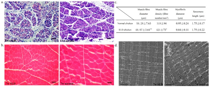
Figure 1.
Muscle fiber characteristics in the sex-linked dwarf (SLD) and normal chickens. (a) Haematoxylin and eosin (H&E) staining of the leg muscle cross-section from embryo day 14 (E14) SLD (left) and normal (right) chickens. Bar, 50 µm; (b) H&E staining of the leg muscle cross-section from seven weeks (7w) SLD (left) and normal (right) chickens. Bar, 50 µm; (c) Muscle fiber diameter, muscle fiber density, myofibrils diameter and sarcomere length in 7w dwarf and normal chickens. Statistical significance between groups was analyzed by one-sample t tests. * p < 0.05; ** p < 0.01; (d) Electron micrograph of a leg muscle cross-section from 7w SLD (left) and normal (right) chickens. Bar, 2 µm.

