Figure 5.
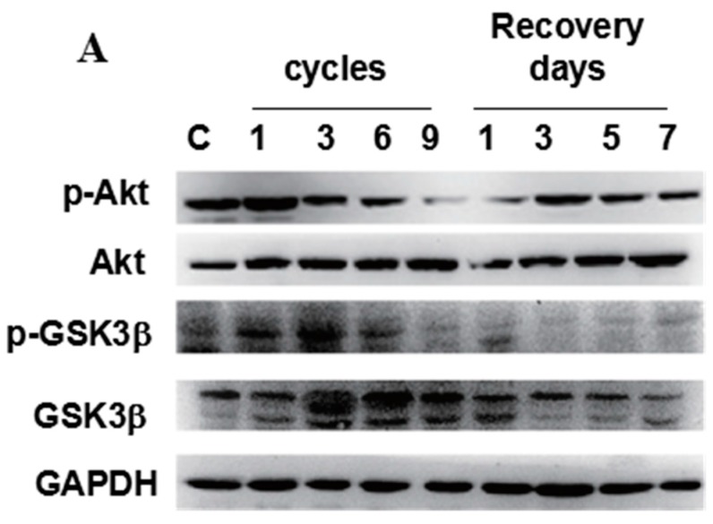
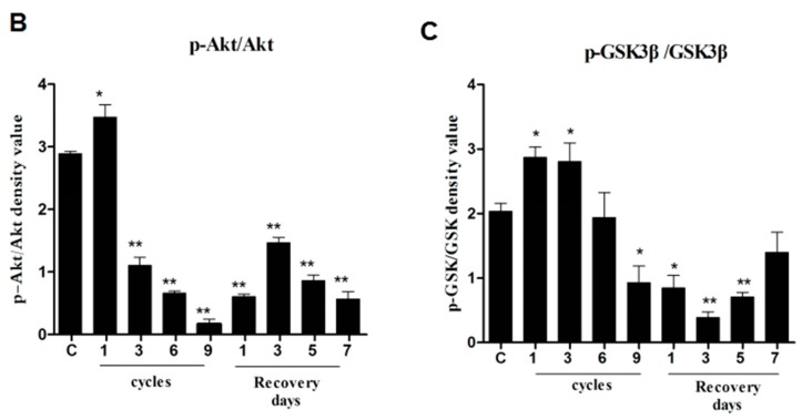
Phospho-Akt and phospho-GSK3β were downregulated in compressed tissues. The protein level of phospho-Akt (p-Akt), Akt, phospho-GSK3β (p-GSK3β), and GSK3β were determined by western blotting. (A) GAPDH was used as a protein loading control and for band density normalization; (B,C) Bar diagram of p-Akt/Akt and p-GSK3β/GSK3β ratios was calculated and plotted. Data are represented as the mean values ± SD. n = 8. * p < 0.05; ** p < 0.01 as compared with the control group (con).


