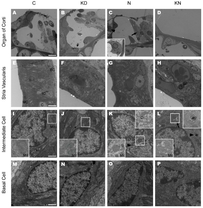
Figure 5.
Ultrastructure of cochleae at the basal turn in control, KD, noise and KN groups. The ultrastructure of the basal turn of the cochlea was observed in control and different experimental groups at P45 (n = 3 in each group). Panels A, B, C and D show the basal OC in control, KD, noise and KN groups. Distinct OHC loss was observed in noise group (C) and the basal OC was collapsed in KN group (D). The inset (panel C) in the left bottom show the inner hair cell; Panels E, F, G and H show the SV at the basal turn in different groups; Panels I, J, K and L show the ultrastructure of IC at the basal turn in control and experimental groups. The insets (panels I, J, K and L) on the left bottom show mitochondria and the myelinbody was magnified in the upper right boxes (Panel K and L). Panels M, N, O and P show the ultrastructure of BC in stria vascularis. Abbreviations: SV: striavascularis; OC: organ of Corti; IC: intermediate cell; BC: basal cell; MC: marginal cell; C: capillary. The scales in panel A, E, I and M represent 10, 5, 1 and 1 μm, respectively. The arrow in L is pointing out a mitochondria and the arrowhead indicates a myelinbody.

