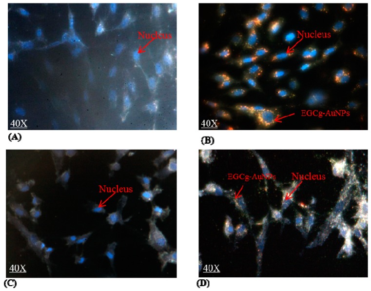
Figure 2.
Cellular internalization of EGCg gold nanoparticles; images were captured via Cyto Viva dark field microscope. (A) human umbilical vein endothelial cells (HUVECs) untreated; (B) HUVECs treated with EGCg-AuNPs 20 μg/mL; 2 h; (C) human coronary artery smooth muscles cells (HCASMCs) untreated; (D) HCASMCs treated with EGCg-AuNPs 20 μg/mL; 2 h. HUVECs: Human umbilical vein endothelial cells, HCASMCs: Human coronary artery smooth muscle cells. Images were captured at 40× magnification.

