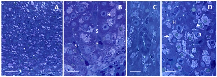
Figure 1.
Light micrographs showing Danio rerio liver under control conditions. (A) Homogeneous organization of liver parenchyma. Bar 25 μm; (B) Hepatocytes (H) surrounding the sinusoids (S) are disposed in cords. The space of Disse (asterisk) lies between the hepatocytes and the sinusoidal endothelium. Bar 10 μm; (C) Bile duct enclosed by cuboidal epithelium. Bar 10 μm; (D) Hepatocytes contain several (arrowhead) lipid droplets surrounded by numerous glycogen granules (arrow). n = nucleus. Bar 10 μm. All toluidine blue.

