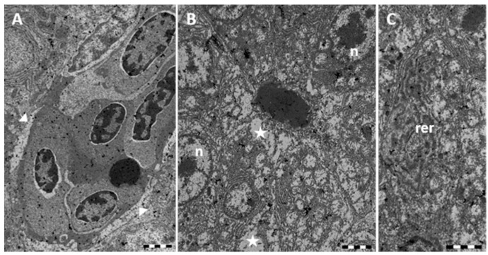
Figure 4.
TEM micrographs of Danio rerio liver section after 96 h of exposure to 7.7 μg/L HgCl2. (A) Alterations in the Disse’s space (arrowheads); (B) In the hepatocyte large cytoplasmic lacunae (star) are detected; nuclei (n) appear damaged; (C) The rough endoplasmic reticulum (rer) is considerably dilated and fragmented. Bar 2 μm.

