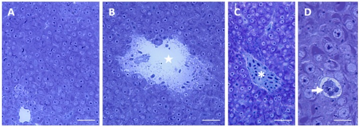
Figure 5.
Light micrographs of Danio rerio liver section after 96 h of exposure to 38.5 μg/L HgCl2. (A) General appearance of liver morphology after 96 h of exposure to 38.5 μg/L HgCl2. Bar 25 μm; (B) Big lysate area (star) in the liver parenchyma. Bar 25 μm; (C) Some blood vessels are occluded (asterisk). Bar 25 μm; (D) Degenerating cell (arrow). Bar 10 μm. All toluidine blue.

