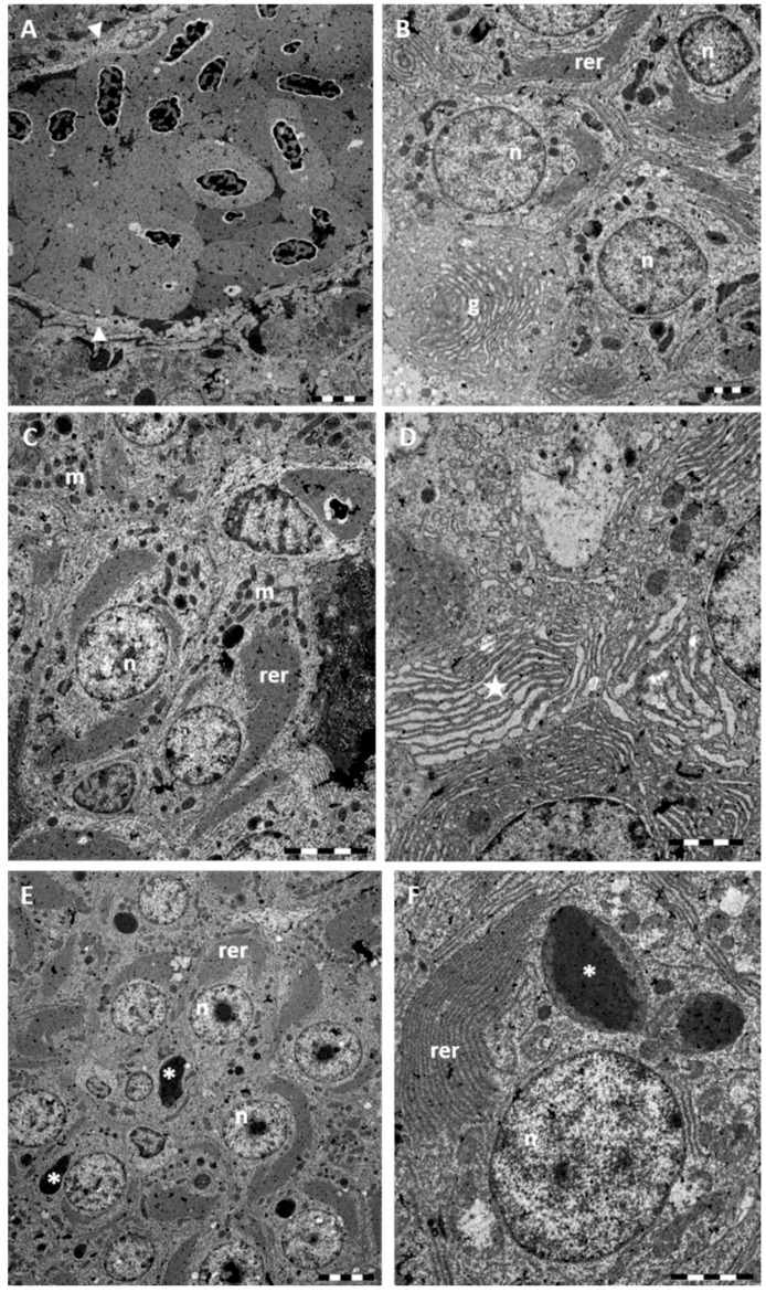
Figure 6.
TEM micrographs of Danio rerio liver section after 96 h of exposure to 38.5 μg/L HgCl2. (A) Occlusion of blood vessel and damaged Disse’s space (arrowheads). Bar 5 μm; (B) RER appear conspicuous and fills most part of the cytoplasm. Note the well-developed Golgi apparatus (g). Some degenerate hepatocyte with pyknotic nuclei can be seen. Bar 2 μm; (C) Numerous mitochondria (m) are scattered through the cytoplasm. Bar 5 μm; (D) High magnification of RER; note at several points, swelling of cisternae (star). Bar 2 μm; (E) In the central portion of nuclei a large nucleolus is evident. In the cytoplasm we can observe the abundant RER and the presence of large electron-dense granules (asterisk) of variable dimension. Bar 5 μm; (F) High magnification of atypical granules (asterisk). Bar 2 μm. n = nucleus; rer = rough endoplasmic reticulum.

