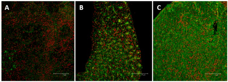
Figure 8.
Confocal micrographs of Danio rerio liver sections labeled with a mouse monoclonal antibody against MT (green—FITC labeled); nuclei labeled with propidium iodide (red). (A) Control samples revealed no or weak signal in few hepatocytes; (B) After 96 h of exposure to 7.7 μg/L of HgCl2, MT immunoreactivity could be observed in hepatocytes cytoplasm; (C) After 96 h of exposure to 38.5 μg/L of HgCl2 the intensity of staining increased and signal was mainly detected in damaged areas. All bar 75 μm.

