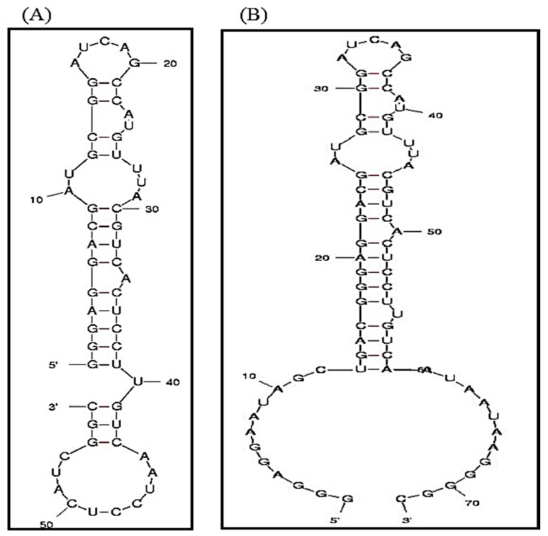
Figure 1.
The secondary structures of (A) A10-3 and (B) A10-3-J1 aptamer, as predicted by the M-fold model. The highlighted region is the sites that are responsible for prostate-specific membrane antigen (PMSA) specificity. This structure is conserved in the A10-3-J1 aptamer to retain the functionality of the aptamer while providing the capability for DNA-RNA hybridization.

