Figure 3.

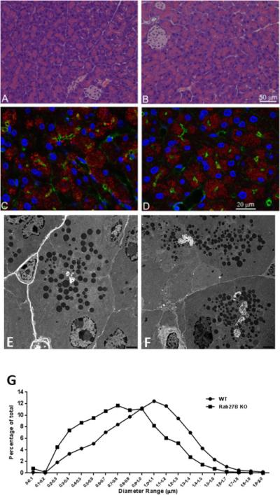
Rab27B deficiency did not affect the general morphology of pancreatic acinar cells and the localization of amylase, but decreased the size of zymogen granules. (A and B) Hematoxylin and Eosin staining was performed on paraffin sections from WT (A) or Rab27B KO (B) mouse pancreas. For immunofluorescence, WT(C) or Rab27B KO (D) pancreas tissues were cryosectioned at 10 μm, stained with phalloidin to label actin (green), DAPI to label nuclei (blue) and anti-amylase antibody (red), respectively. Images were obtained using confocal microscopy. (E and F) Pancreas tissues from WT (E) and Rab27B KO (F) mice were processed for electron microscopy as described in Material and Methods. Images were obtained at the magnification of 6000X. (G) The diameter of zymogen granules from WT and KO mouse pancreas was measured using images obtained from electron microscopy. Each data set represents a combination of values obtained from 7-12 sections from each of three mice. The values were normalized to the total number of granules.
