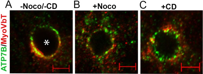
Fig. 4.

Microtubule and actin disruption completely disperses apical MyoVbT, but partly disperses apical ATP7B. (A) In cells not treated with nocadazole and/or cytocalasin D (−Noco/−CD) ATP7B (green) and MyoVbT (red) colocalize near the bile canaliculus. (B) Disruption of microtubules with Nocodazole (+Noco) disperses MyoVbT to numerous peripheral vesicles. ATP7B is also dispersed into vesicles following Nocodazole treatment, and a Nocodazole-insensitive pool remains associated on the apical membrane. (C) Disruption of actin polymerization with cyotchalasin D (+CD) also disperses ATP7B and MyoVbT into a discreet vesicular pool. This pool of vesicles is associated with the cortical actin adjacent to the bile canaliculus. Asterisk indicates a bile canaliculus. Scale bars: 2 µM.
