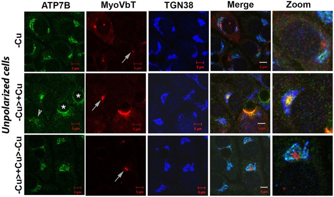
Fig. 6.
MyoVbT arrests ATP7B in the apical compartment of unpolarized cells in high [Cu+]. WIF-B cells were transformed with virus on day 11 in culture and fixed 16 h later; ∼10% of the cells remained unpolarized. MyoVbT (red) localizes to the apical compartment under all Cu+ conditions (white arrows, second column). ATP7B (green) colocalizes with TGN38 (blue) in Cu+ depleted conditions and shows no overlap with MyoVbT in the apical compartment (top panel). Following Cu+ treatment, ATP7B traffics from the TGN and colocalizes with MyoVbT in the apical compartment (middle), and at the bile canaliculus in polarized cells (asterisks). In cell not expressing MyoVbT, ATP7B disperses to vesicles (arrowhead). On subsequent Cu+ chelation, ATP7B exhibits retrograde trafficking denoted by its overlap with TGN38 (bottom). Scale bars: 5 μm. Details of the boxed areas are magnified in the three right panels, showing the arrangement of ATP7B, MyoVbT, and TGN38 near the apical compartment.

