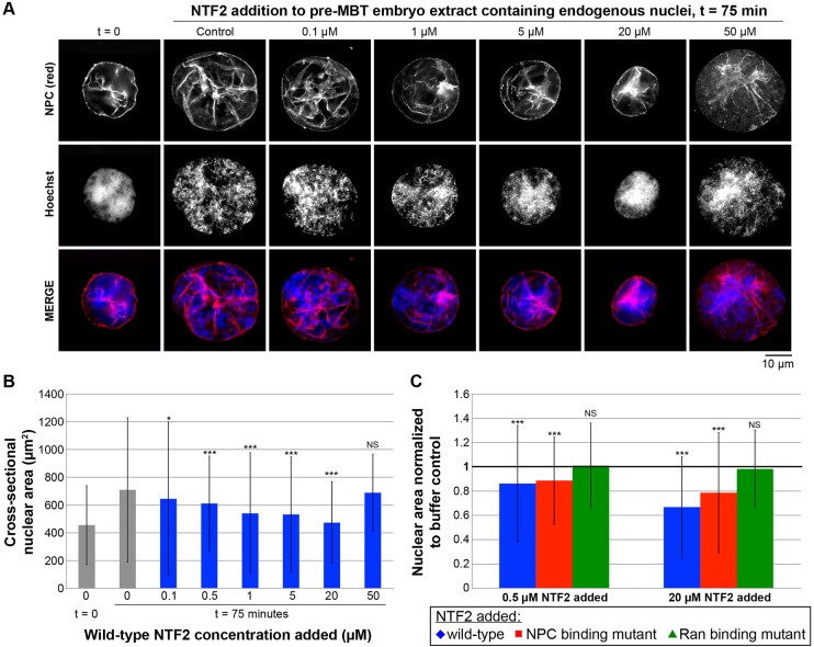
Fig. 3.
NTF2 concentration affects nuclear size in X. laevis embryo extracts. (A) Embryo extracts containing endogenous embryonic nuclei were prepared from X. laevis embryos at early stage 8. Recombinant NTF2 protein was added to the indicated final concentrations, and the same total volume was added to each reaction. For the control reaction, an equal volume of NTF2 storage buffer was added. Nuclei were incubated for 75 min at 20°C, during which time some nuclear growth occurs. Nuclei were then fixed, spun onto coverslips, processed for immunofluorescence, and stained with mAb414 to visualize the NPC (red) and Hoechst 33342 to visualize the DNA (blue). Representative images are shown. (B) For each sample, the cross-sectional areas of 127–625 nuclei were measured from NPC-stained nuclei and averaged. Average nuclear size data from three different embryo extracts are shown. The error bars are s.d. For the statistical analysis, calculations were performed relative to the no-NTF2-added control at t=75 min. (C) Average cross-sectional nuclear areas were measured as in B and normalized to the buffer-addition control (bold horizontal line set at 1.0). Average nuclear size data from three different embryo extracts are shown. The error bars are s.d. For the statistical analysis, calculations were performed relative to the no-NTF2-added control at t=75 min. *P<0.05; ***P<0.001; NS, not significant (two-tailed Student's t-test).

