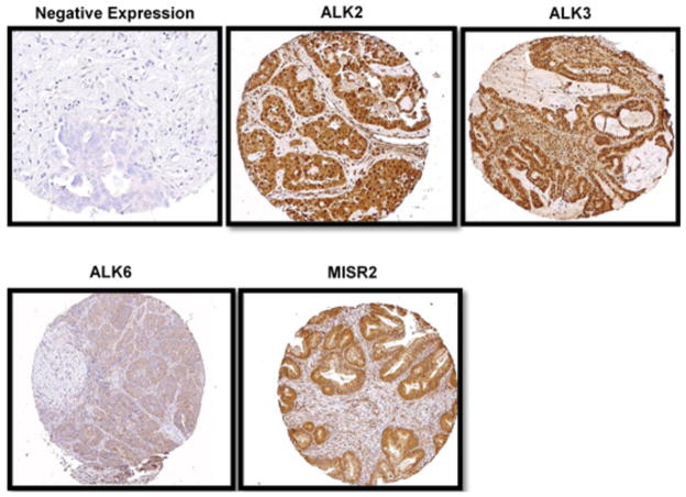
Fig. (1).
Immunohistochemical staining of tissue microarray of epithelial ovarian cancer samples. Representative images depict negative and positive staining for ALK2, ALK3, ALK6 and MISR2 in a TMA core. TMAs were created from formalin-fixed, paraffin-embedded tumors and stained with antibodies to four proteins of interest:, ALK2, ALK3, ALK6 and 12G4mab. Digital images of each core were captured and stored using a high resolution microscope (Zeiss Axioplan, CA) and video camera interfaced with Microsoft access software to correlate each tissue core with the appropriate patient identifier. Digital images of the stained specimens were reviewed and scored independently by two reviewers, blinded to clinical information.

