Fig. 1.
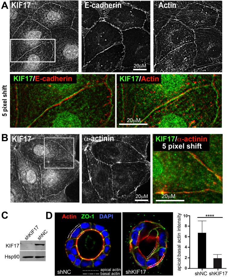
KIF17 localizes at cell–cell junctions and contributes to actin remodeling during epithelial morphogenesis. (A) Colocalization of KIF17 with actin and E-cadherin at cell–cell contacts in MDCK cells. Color overlay shows an enlarged view of the boxed region of KIF17 and E-cadherin or β-actin images; the KIF17 image was shifted by five pixels to highlight corresponding staining patterns. (B) Colocalization of KIF17 and α-actinin at cell–cell junctions. Color overlay shows an enlarged view of the boxed region; the KIF17 image was shifted by five pixels. (C) Immunoblot showing KIF17 in MDCK cells transduced with control (shNC) or KIF17-targeting (shKIF17) shRNAs. Hsp90 was used as a loading control. (D) Localization of actin (phalloidin), ZO-1 and nuclei (DAPI) in shNC and shKIF17 MDCK cysts grown in Matrigel for seven days. Dotted and dashed lines highlight apical and basal membranes, respectively. Graph shows the ratio of apical to basal actin fluorescence intensity determined by line-scan analysis. n=38 and 48 cells for shNC and shKIF17, respectively. Error bars are s.e.m., significance was determined with a two-tailed, unpaired student's t-test, ****P<0.0001.

