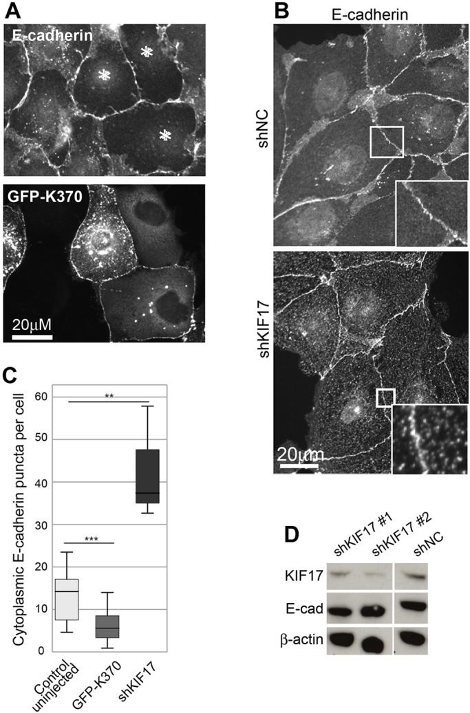
Fig. 7.

KIF17 regulates the distribution of E-cadherin. (A) Localization of E-cadherin in MDCK cells expressing GFP–K370 or GFP–KIF17-FL and fixed 4 h after cDNA injection. Asterisks mark injected cells. (B) Localization of E-cadherin in cells transduced with shNC or shKIF17. Boxed regions are magnified in insets. (C) Box-whisker plots showing quantification of cytoplasmic E-cadherin puncta in uninjected controls and in cells expressing GFP–K370 and in KIF17-depleted cells (shKIF17). Results are from images of injected cells in 2–4 independent experiments. Significance was determined using a two-tailed Mann–Whitney U test. **P<0.01; ***P<0.001. (D) Immunoblots showing KIF17, E-cadherin and β-actin in MDCK cells transduced with shNC, shKIF17#1 or shKIF17#2.
