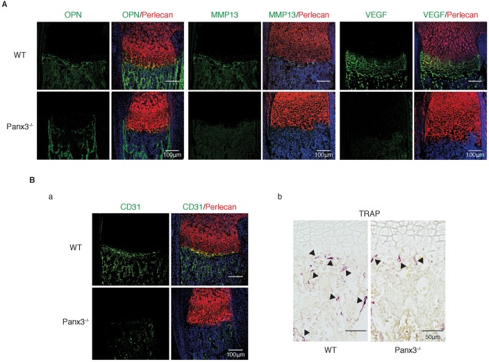
Fig. 3.
Panx3 knockout inhibits chondrocyte terminal differentiation. (A) Immunostaining of the growth plate of newborn WT and Panx3−/− tibias with antibodies to OPN (green), MMP13 (green) or VEGF (green). (B) CD31 (green) staining (a) and TRAP staining (b) of the growth plate of WT and Panx3−/− tibias. Perlecan (red) staining shows cartilage areas; nuclei were stained with Hoechst dye 33342 (blue in A and Ba).

