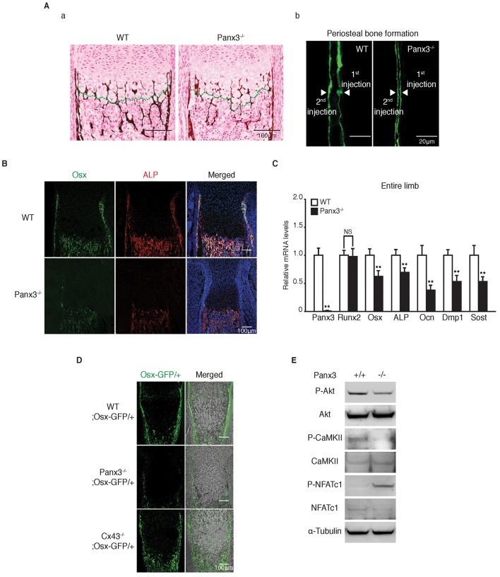
Fig. 4.
Panx3 knockout inhibits osteoblast differentiation. (A) (a) Double staining of Safranin-O (red) and von Kossa (brown) of the growth plates of tibias in newborn WT and Panx3−/− mice. (b) New bone formation was analyzed by double calcein labeling [injecting calcein at P10 (1st injection) and at P17 (2nd injection)]. Samples were collected at P18. The spacing of the calcein-labeled layers between the 1st injection and the 2nd injection, shown by arrowheads, represents the bone formed between the injection times. (B) Immunostaining of the growth plates in tibias of newborn WT and Panx3−/− mice for Osx (green) and ALP (red). The nuclei were stained with Hoechst 33342 (blue). (C) qPCR for Panx3, osteoblast markers (Runx2, Osx, ALP and Ocn) and osteocyte markers (Dmp1 and Sost) using total RNA prepared from whole tibias of newborn mice. Results represent the mean±s.d., n=3. **P<0.01. NS, not significant (Student's t-tests). (D) Osx–GFP expression in Panx3−/− and Cx43−/− growth plates. The Osx–GFP expression is reduced in the Panx3−/− growth plate but not the Cx43−/− growth plate. Osx–GFP expression of the growth plates in the tibias of newborn WT Osx-GFP/+ mice, Panx3−/−;Osx-GFP/+ mice, and Cx43−/−; Osx-GFP/+ mice. The right panels show merged images of GFP (green) and light microscopy images. (E) Western blots of signaling molecules in WT and Panx3−/− calvarial cells. The Panx3 ER Ca2+ channel promotes NFATc1 activity. Downregulation of activities of signaling molecules in the Panx3 ER-mediated NFATc1 pathway in Panx3−/− calvarial cells. Calvarial cells were prepared from newborn WT and Panx3−/− mice and phosphorylation levels (P-) of Akt, CaMKII, and NFATc1 were analyzed by western blotting.

