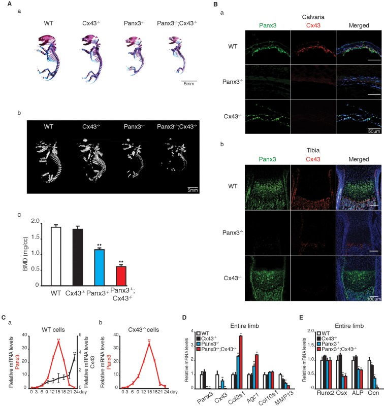
Fig. 6.
Panx3 is upstream of Cx43 in osteoblast differentiation. (A) (a) Alizarin Red and Alcian Blue staining of the whole skeletons of newborn WT, Cx43−/−, Panx3−/− and Panx3−/−;Cx43−/− mice. (b) μCT analysis of WT, Cx43−/−, Panx3−/− and Panx3−/−;Cx43−/− bones. (c) Quantification of bone mineral density (BMD). (B) Immunostaining. (a) The newborn calvarias (a) and growth plates of the tibias (b) were stained with antibodies to Panx3 (green) and Cx43 (red). The nuclei were stained with Hoechst dye 33342 (blue). (C) qPCR analyses of Panx3 and Cx43 mRNA expression during osteoblast differentiation in primary calvarial cells from WT (a) and Cx43−/− mice (b). Primary calvarial cells were cultured in the osteogenic induction medium. Total RNA was extracted from the cells at the indicated days after the osteogenic induction. (D) qPCR analysis of the mRNA expression of the chondrogenic marker genes and the Panx3 and Cx43 genes using RNA isolated from the whole tibias of newborn WT, Cx43−/−, Panx3−/− and Panx3−/−;Cx43−/− mice. (E) qPCR analysis of the mRNA expression of osteogenic marker genes using RNA isolated from the whole tibias of newborn WT, Cx43−/−, Panx3−/−, and Panx3−/−;Cx43−/− mice. Results represent the mean±s.d., n=3. *P<0.05, **P<0.01 [Student's t-test (Ca,Cb) and one-way ANOVA (Ac,D,E)].

