Fig. 2.
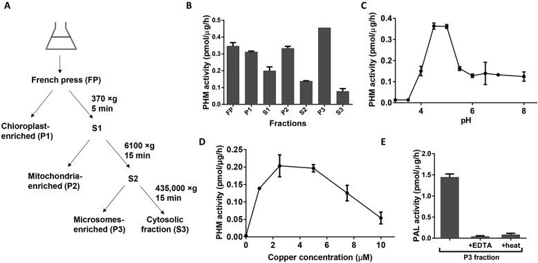
Chlamydomonas cell lysates contain active PAM. (A) Schematic of subcellular fractionation protocol for Chlamydomonas. (B) PHM activity was detected in all fractions. (C) Microsomal PHM activity plotted as a function of pH. (D) Microsomal PHM activity was greatest at 2.5–5 µM copper. (E) The microsomal fraction displayed PAL activity that was inhibited by a divalent metal ion chelator (EDTA) or heat (5 min at 100°C). Data shown are mean±s.d. (n=2 or 3). Graphs are from one of two experiments, which yielded similar results.

