Fig. 4.
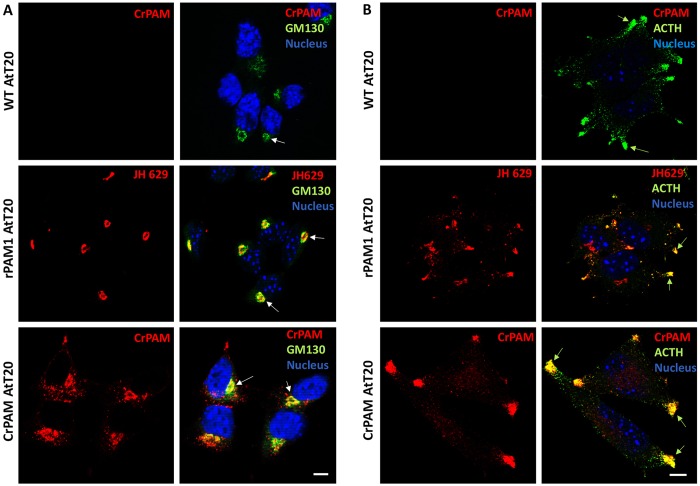
Expression of CrPAM in mammalian neuroendocrine cells. Wild-type AtT-20 cells (WT) and stable lines expressing rPAM1 or CrPAM were stained for GM130 (A), a Golgi marker, or ACTH (B), a granule marker, using a FITC-tagged secondary antibody; cells were pretreated with 10 µM cycloheximide for 1 h (Sobota et al., 2006). WT and CrPAM-expressing cells were stained using an affinity-purified anti-CrPAM C-terminal antibody; no background was observed in WT AtT-20 cells (upper panel, A and B). rPAM1 AtT-20 cells were stained with antibody to the linker region of mammalian PAM (JH629). PAM staining was visualized using a Cy3-tagged secondary antibody. (A) Optical sections through the Golgi area showed perinuclear staining in rPAM1- and CrPAM-expressing cells that partially colocalized with GM130 (white arrows). (B) In optical sections near the bottom of each cell, rPAM1 and CrPAM were identified at the tips of cellular processes, where ACTH-containing granules accumulated (green arrows). Scale bars: 10 µm. Nuclei were visualized with Hoechst 33342 stain.

