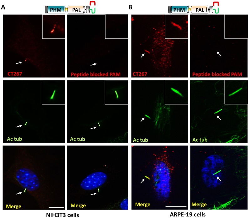
Fig. 8.
PAM localizes to sensory cilia in mammalian cells. (A) Left, serum-starved NIH3T3 cells immunostained with antibody to acetylated tubulin and the C-terminus of mammalian PAM (CT267) displayed PAM staining along the length of the cilium (white arrow points to the cilium). Scale bar: 10 µm. Right, PAM protein could not be detected when the antibody was preincubated with the antigenic peptide. (B) Left, human retinal pigment epithelial cells immunostained with antibodies to the C-terminus of mammalian PAM (CT267) and acetylated tubulin (Ac tub) displayed a similar localization of PAM in the cilium (white arrow points to the cilium). Right, peptide blocking decreased the signal observed in the cilium. Scale bar: 10 µm.

