
Fig. 5.
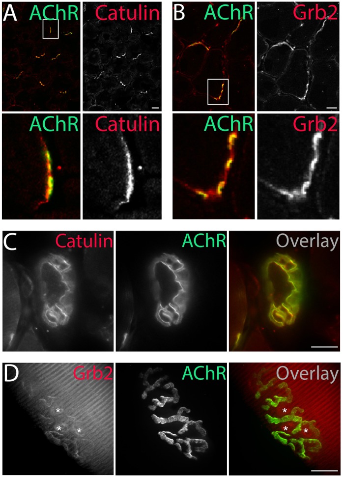
Localization of α-dystrobrevin-1-interacting proteins to the NMJ. α-Catulin (A) and Grb2 (B) (antibodies, red) concentrate at the postsynaptic machinery of NMJs (AChR, green), as visualized by immunohistochemical analysis of 6-μm-thick slices of tibialis anterior muscle. Immunostaining of α-catulin in the triangularis sternum muscle (C) and Grb2–GFP fluorescence in the tibialis anterior muscle (D) (both shown in red) mirrored the localization of AChRs (green). Asterisks in D indicate fluorescence at synaptic nuclei (see also Fig. 6B). Scale bars: 20 μm.