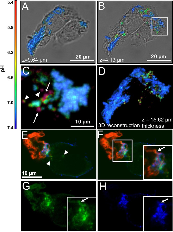
Fig. 1.

AgLDL is sequestered in compartments that are surface-connected and contain acidified sub-regions where free cholesterol is generated. (A–D) Ratiometric confocal images of J774 cells incubated with agLDL dual-labeled with a pH-sensitive and pH-insensitive probe for 1 h. (A) Aggregate contained above the surface of the cells and (B) agLDL in the middle of the cells. (C) Enlargement of an area containing acidified regions, highlighted by a box in B. Regions of acidification that represent completely internalized vesicles (arrowheads) and areas of low pH that retain their connection to portions of the aggregate that are at neutral pH (arrows) can be seen. (D) 3D reconstruction of the data shown in A and B. (E–H) RAW264.7 macrophage-like cells were incubated with Alexa633–agLDL (red) for 1 h, stained with Alexa555–CtB (blue) on ice to label the plasma membrane, fixed and stained with filipin (green) to visualize free cholesterol. (E) A cell is contacting Alexa633–agLDL. Some filipin labeling of free cholesterol can be seen inside the cell (arrowheads). (F–H) An extracellular strand of agLDL in close proximity to the cells is shown by an arrow in the inset (F). Single color images of the same region show that it is labeled with filipin (G), but the cell only touches this strand at the base (H). There are areas of filipin labeling that are also labeled with CtB, suggesting that these are also associated with the plasma membrane.
