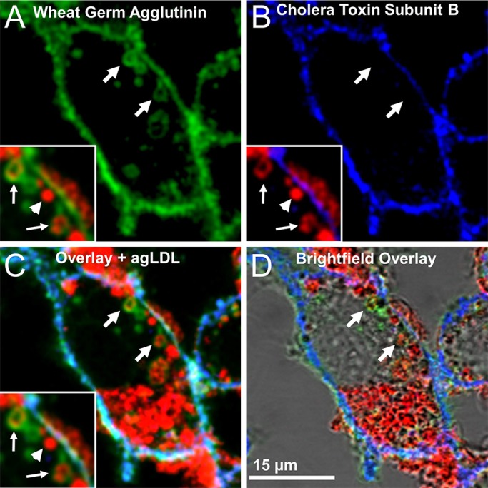
Fig. 3.

AgLDL resides in sub-compartments that retain surface connectivity but have restricted access to the extracellular space. J774 cells were incubated with Alexa633–agLDL for 1 h, stained with Alexa555–CtB on ice for 5 min, fixed with 3% PFA and 0.5% glutaraldehyde to block the diffusion of lipid-linked proteins (CtB) following fixation and then labeled with Alexa488–WGA. (A) WGA staining shows compartments (arrows) that contain agLDL (red, inset) and are surface connected. Some regions of agLDL represent completely internalized vesicles that are negative for WGA (arrowhead, inset). (B) CtB staining labels the surface of the cell but was largely excluded from areas of agLDL sequestration (blue, arrows, inset). (C) Overlay and (D) brightfield overlay of WGA staining, CtB staining and agLDL, showing that agLDL resides in surface-connected compartments that seal sufficiently to exclude CtB (C, arrows, inset).
