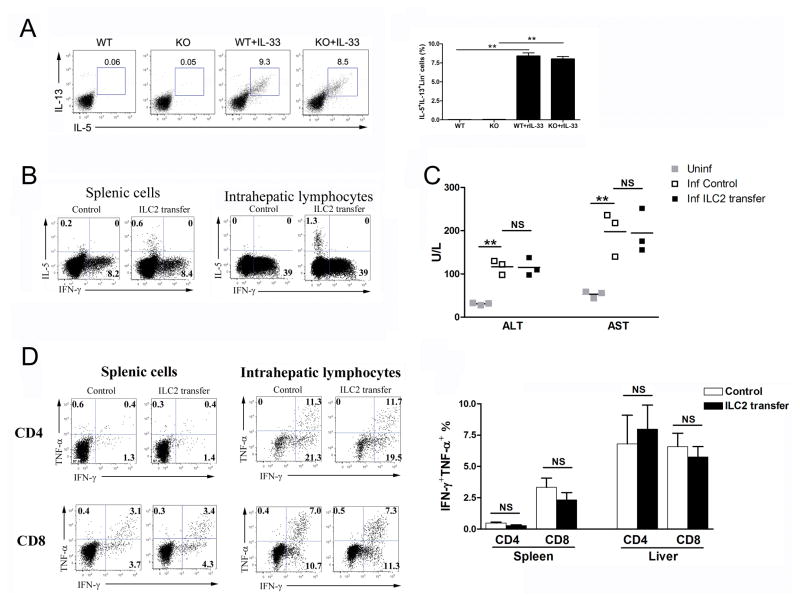
Figure 5. ILC2s are dispensable for anti-viral T-cell responses.
(A) WT and IL-33−/− mice were infected, followed by rIL-33 treatment (1 μg/mouse, PBS was used as a control) daily starting from 1 dpi. All mice were sacrificed at 7 dpi. IHLs were stimulated with PMA/Iono for 4 h in the presence of GolgiStop, followed by the surface staining of lineage markers (Lin) and intracellular IL-5 and IL-13. Lin- cells were gated out first and further analyzed for IL-5+IL-13+ populations. (B) 5×10⁶ ILC2s were adoptively transferred into LCMV-infected WT mice at 1, 3 and 5 dpi (PBS was used as a control). Total live cells from spleens and livers were gated for detection of IL-5- and IFN-γ-producing cells. (C) Serum ALT and AST levels were evaluated by enzymatic reaction assay. Symbols represent individual mice and bars represent means. (D) Splenic and hepatic virus-specific T-cell responses were evaluated by intracellular IFN-γ and TNF-α staining. Data are shown as mean + SEM of 3–5 mice/group, from single experiments representative of at least three experiments performed. A two-tailed t test was used to compare the two groups. * P<0.05, ** P<0.01.

