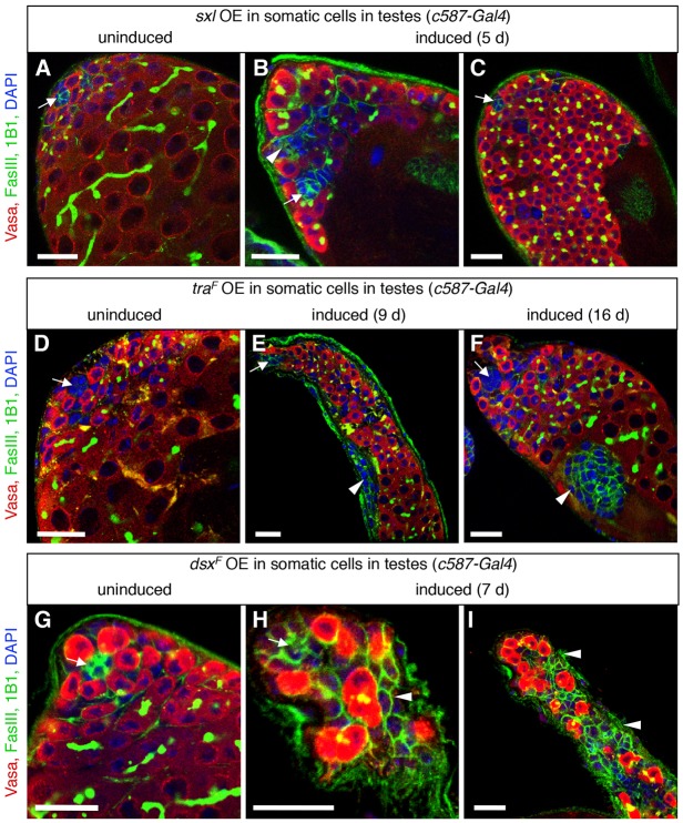
Fig. 4.
Ectopic expression of female sex determinants in the adult testis disrupts testis morphology. (A-I) Immunofluorescence detection of FasIII (green at cell periphery) and 1B1 (green in germ cell fusomes) to visualize testis morphology before or after ectopic expression of female sex determinants in cyst stem cells and early cyst cells in adult testes. Vasa (red) marks germ cells; DAPI (blue) marks nuclei; arrows mark the hub. Before expression of sxl (A), traF (D) or dsxF (G), testes look normal. After ectopic overexpression (OE) of sxl (B,C) or traF (E,F), aggregates of FasIII+ somatic cells (arrowheads) and overproliferating early germ cells accumulate at the testis apex. After ectopic expression of dsxF (H,I), germ cells fail to differentiate and aggregates of somatic cells accumulate at the testis apex. Scale bars: 20 μm.

