Figure 7.

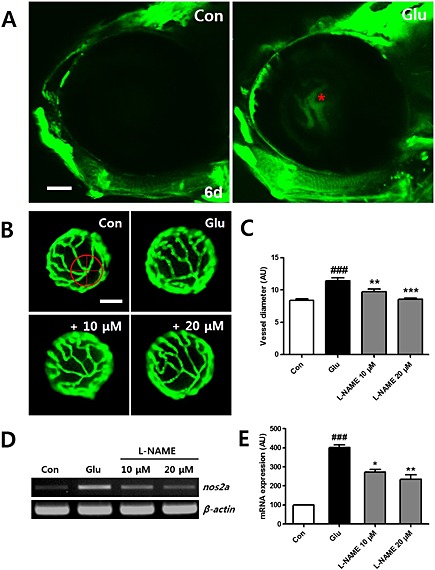
NO production by HG in hyaloid‐retinal vessels. (A) Comparison of NO production using a DAF‐FA DA probe in hyaloid‐retinal vessels in control larvae at 6 dpf. NO production is not observed in control larvae but is detected in HG‐treated larvae (red asterisk). Scale bar = 50 µm. (B) The effect of the NO inhibitor on dilated hyaloid‐retinal vessels. HG‐induced vessel dilation was rescued by the NOS inhibitor (l‐NAME, 10 and 20 μM). (C) Graph data are displayed as the mean AU for vessel diameter. The vessel diameter of each lens was measured three times. Scale bar = 40 µm. ###P < 0.001 versus control; **P < 0.01, ***P < 0.001, significantly different from HG, n = 10 embryos per group. [(D) and (E)] Expression of nos2a in HG‐treated larvae increased significantly compared to control and reduced by treatment with l‐NAME. No changes in the transcript levels of β‐actin were detected between control and HG‐treated larvae. ###P < 0.001, significantly different from control; *P < 0.05, **P < 0.01, significantly different from HG.
