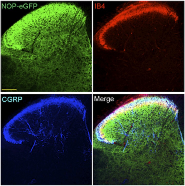
Fig. 4.
NOP-eGFP receptors are highly distributed in laminae I-III and ×. Tissue sections from the spinal cord were incubated with anti-GFP, and –CGRP (laminae I and IIo, panel A). Tissues were also treated with biotinylated IB4 (dorsal border of lamina IIi) and streptavidin. This figure is reprinted with permission from the Journal of Neuroscience.

