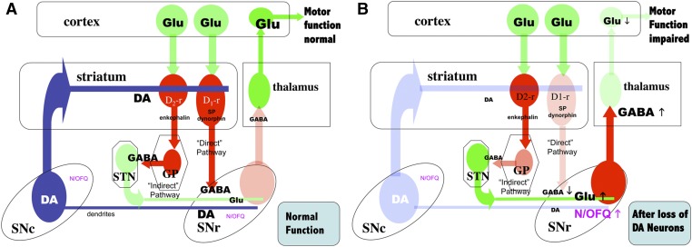
Fig. 7.
Schematic representation of the organization of basal ganglia regulating motor function and the effects dopamine (DA) depletion on N/OFQ expression and release. DA neurons are represented in blue; glutamate (Glu) neurons in green; GABA neurons in red; color density indicates the relative levels of activity in each system with normal DA neuron function (Panel A, normal function), or after loss of a significant fraction of DA neurons (Panel B). GP, globus pallidus; N/OFQ, nociception/ orphanin FQ; SNc, substantia nigra compacta; SNr, substantia nigra reticulate; STN, subthalamic nucleus, Panel A. With DA neuron function intact, GABA release in the pallido-subthalamic neurons in the “indirect” striato-nigral pathway reduces Glu release from the subthalamic neurons that activate the GABAergic nigrothalamic pathway. With low release of GABA in the thalamus from this pathway, the thalamocortical glutamatergic neurons are active, increasing activity in motor cortex and maintaining normal motor function. N/OFQ levels and release in the SNr are relatively low under these conditions. Panel B. When nigrostriatal DA function is impaired (e.g., after 6-OHDA or MPTP treatment), activity in the subthalamic glutamatergic neurons to the SNr is increased, resulting in activation of the nigrothalamic GABA pathway and inhibition of thalamocortical neurons that facilitate normal motor function. After 6-OHDA or MPTP treatment, ppN/OFQ mRNA and N/OFQ levels and release in SNr are increased (Marti et al., 2005, 2010); N/OFQ release in SNr is also increased by haloperidol treatment (Marti et al., 2010). NOPr antagonists largely reverse the effects of DA depletion by 6-OHDA on GABA release in SNr and thalamus (Marti et al., 2005, 2007, 2010). Treatment with 6-OHDA also reduces NOPr mRNA expression in SNc (Norton et al., 2002; Marti et al., 2005). Arrows indicate the direction of change in N/OFQ, GABA or Glu release after 6-OHDA or MPTP treatment.

