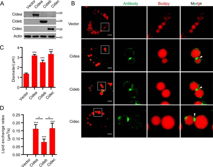
FIGURE 1.
CIDE proteins promote LD fusion and large LD formation in HepG2 cells. A, Vector, Cidea, Cideb, and Cidec plasmids were transfected into HepG2 cells. Expression levels of CIDEs were detected by Western blot. B, protein localization of CIDEs in HepG2 cells as in A. Oleic acid was added to promote the formation of LDs for 15 h. LDs were labeled with Bodipy 556/568 (C12, red). Scale bars, 10 μm. Arrowheads point to LDCSs. C, largest lipid droplet size per cell was measured in A. Ten cells were analyzed in each group. D, lipid exchange rates were measured in A. Five pairs of LDs were measured. Quantitative data are presented as the mean ± S.E. Differences were considered significant at p < 0.05. *, p < 0.05; ***, p < 0.001.

