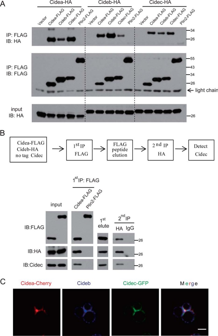
FIGURE 3.
CIDE proteins interact with each other and form a complex at LDCSs. A, FLAG-tagged CIDEs and Plin2 together with HA-tagged CIDEs were coexpressed in 293T cells. Anti-FLAG M2 beads were used for immunoprecipitation. The immunoprecipitated products were detected by antibodies against FLAG or HA. IP, immunoprecipitation; IB, immunoblot. B, two-step coimmunoprecipitation of complex containing Cidea, Cideb, and Cidec. Top, schematic showing procedures for two-step coimmunoprecipitation assay. Cidea-FLAG or Plin2-FLAG (as a control) was transfected into 293T cells with Cideb-HA and untagged Cidec. C, Cidea, Cideb, and Cidec were colocalized at the LDCS. HepG2 cells were cotransfected with Cidea-Cherry (red), Cidec-GFP (green), and Cideb. Cells were stained with antibodies against Cideb (blue). Scale bar, 2 μm.

