FIGURE 2.
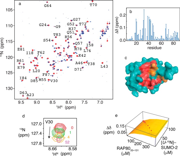
a, two-dimensional 1H-15N HSQC NMR spectra for free SUMO-2 (red) and bound to RAP80-(33–131) (blue). b, chemical shift perturbations for SUMO-2 bound to a 52-fold excess of RAP80-(33–131). c, residues experiencing chemical shift changes greater than 1 S.D. from the mean (red) and those broadened beyond detection (orange) are mapped on the surface of SUMO-2 (PDB code 1WM2). d, expanded region from the two-dimensional 1H-15N HSQC NMR spectra taken during titration of SUMO-2 with RAP80-(33–131), showing chemical shift changes for Val30, with ligand/protein ratios indicated. e, fits of chemical shift perturbation data to 1:1 binding isotherms for the SUMO-2 interaction with RAP80-(33–131). Chemical shift changes for Val30 are indicated on the vertical axis, and concentrations for SUMO-2 and unlabeled RAP80-(33–131) are indicated on the horizontal axes. Experimentally determined chemical shift changes are shown as points, and the best fit to a 1:1 binding isotherm is shown as a surface.

