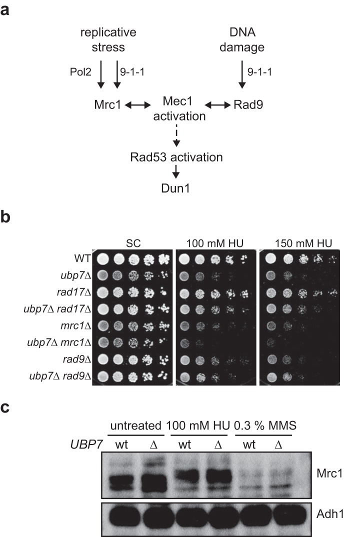
FIGURE 4.

Epistasis analysis of UBP7 with intra-S phase checkpoint genes. a, schematic of relevant factors for Mec1 and Rad53 activation. b, 5-fold serial dilutions of the indicated strains spotted on plates containing HU and incubated at 30 °C for 3 d. c, Mrc1 protein levels and phosphorylation status in wild-type and ubp7Δ MRC1–9myc strains. Cultures were left untreated or exposed to 100 mm HU or 0.3% MMS for 2 h before whole-cell lysates were prepared. Shown is a Western blotting analysis against myc (Mrc1) and Adh1 (loading).
