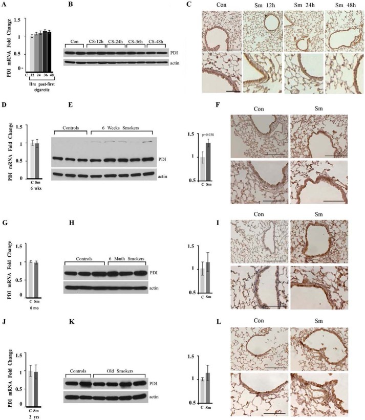
FIGURE 2.
PDI protein is up-regulated in murine smokers. Wild-type C57Bl/6 mice were exposed or not to CS for the indicated periods of times. Lungs were harvested and lysed. Exposure periods are one-time smokers (A–C); 6-week chronic smokers (D–F); 8-month-old chronic smokers, exposed for 6 months (G–I), and 2-year-old chronic smokers, exposed for 6 months (J–L). Amount and distribution of PDI mRNA (A, D, G, and J) and PDI protein (B, E, H, and K) were determined in total lung lysates of control (marked as C or Con) and smoke-exposed (marker as Sm) animals by quantitative RT-PCR and Western blotting analysis. The graph on the right represents quantitation of the amounts of PDI protein by densitometry units (p = 0.038 for 6-week smokers; p = 0.2601 for 8-month smokers; and p = 0.2064 for old smokers). Actin was used as loading control. C, F, I, and J, lungs of age-matched controls (right panels) and smokers (left panels) were stained as one set using identical conditions with anti-PDI antibody. Upper panels images acquired at ×200 magnification. Lower panels images represent enlarged areas from original images acquired at ×400 magnification. Scale bars, 100 μm for the upper panels and 200 μm for the lower panels. Images are representative of at least three animals analyzed for each group.

