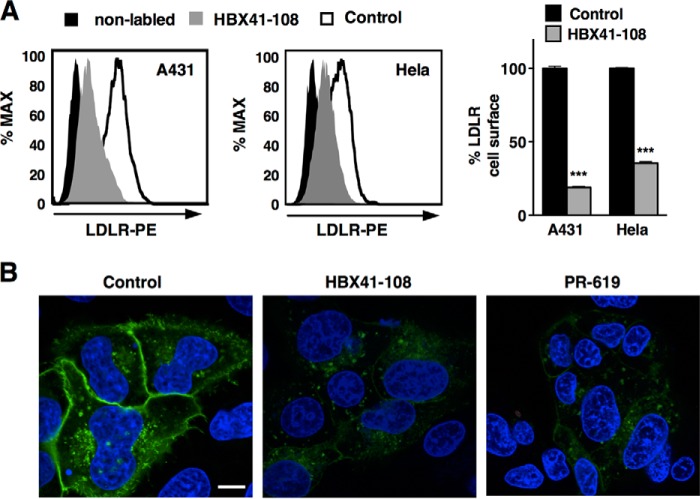
FIGURE 4.
DUB inhibition promotes removal of cell surface LDLR. A, the human A431 and HeLa cell lines were cultured in sterol depletion medium 18 h prior to being treated with 5 μm HBX41-108 for 2 h. Cell surface LDLR was determined by FACS analysis. A representative FACS histogram and the quantification of the experiments are shown (n = 3). B, HepG2-LDLR-GFP cells were treated with vehicle (control), 5 μm HBX41-108, or 50 μm PR-619 for 2 h. Representative images are shown with LDLR in green and staining of nuclei with DAPI in blue. Size bar, 10 μm. ***, p < 0.001.

