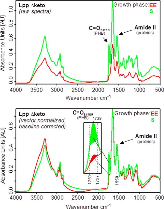
FIGURE 5.

Determination of the relative PHB amounts from whole intact cells of L. pneumophila. Upper panel, original (raw) absorbance spectra of L. pneumophila Paris Δketo (Lpp Δketo). Lower panel, preprocessed absorbance spectra of L. pneumophila Paris Δketo. Preprocessing involved vector normalization in the amide II region (1520–1570 cm−1) and offset correction. The intensity of the ester carbonyl band around 1739 cm−1 of spectra normalized to the amide II band can be used to determine the relative amount of PHB present in the cells. For this purpose, the areas under the curves are calculated between 1727 and 1750 cm−1 (see inset of the lower panel).
