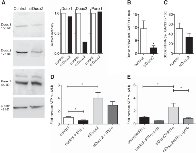
FIGURE 2.
Suppression of Duox2 by lentiviral infection with siDuox2. A, representative Western blotting (pooled from three lung donors) and protein quantification (corrected for β-actin, pooled samples from three donors and run in duplicate) for Duox 1 and 2, pannexin 1 (Panx1), and β-actin after infection with control plasmid or shRNA for Duox2 (see “Experimental Procedures”). Duox2 protein is down-regulated but Duox1 and Panx1 are not. There is a minor band in the panx1 blots. Exclusion or inclusion of this band in the quantification did not make a difference. B and C, mean ± S.E. of mRNA expression (relative to GAPDH × 100) after infection with control plasmid or shRNA for Duox2 (see “Experimental Procedures”). Duox2 mRNA is down-regulated (B, n = 3 lung donors, 3 replicates each) but SOD 2 is not (C, n = 3 lung donors, 3 replicates each). D and E, Duox2 shRNA eliminates the IFN-γ effect on ATP release and leads to an increase in ATP release at baseline. The sustained ATP release in cells expressing siDuox2 can be inhibited by probenecid (E), indicating that this ATP release occurs through Panx1. Data are mean ± S.E. of -fold increases of ATP release (peak release) of control (non-targeting shRNA) or Duox2 shRNA-infected cells ± IFN-γ ± probenecid. n = 3 lung donors, 3 replicates each. *, p < 0.05.

