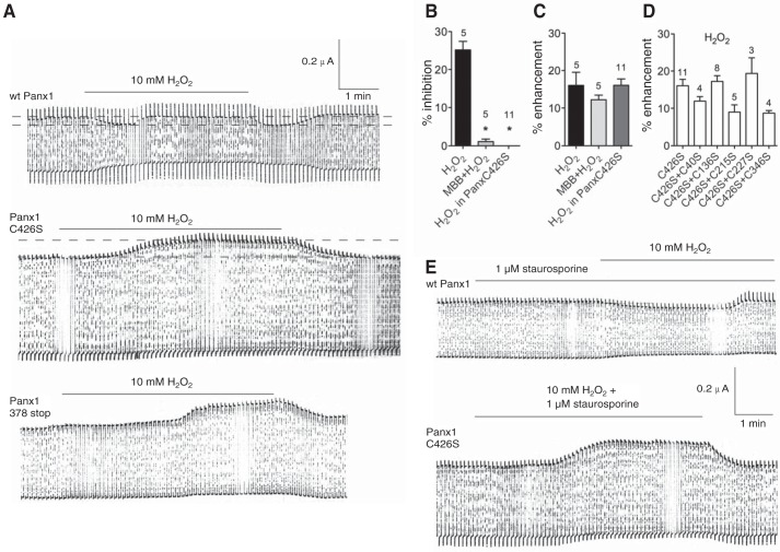
FIGURE 3.
Electrophysiological recordings of membrane currents of an oocyte expressing Panx1. A, membrane currents were measured of oocytes expressing WT Panx1, the replacement mutant Panx1 C426S, or the truncation mutant 378stop. Oocytes were held at −60 mV, and pulses to +10 mV were applied at a rate of 0.1 Hz to transiently open the channels. Application of 10 mm H2O2 resulted in a biphasic response in WT Panx1, where an initial attenuation was followed by recovery to base levels or even a small increase of the currents. Following washout of H2O2, the currents dropped to the level of maximal inhibition but then recovered to the level seen before the interference. With both Panx1 mutants, only an increase in current amplitude was observed that reversed upon washout of H2O2. B and C, quantitative analysis of the inhibitory (B) and enhancing (C) components of the H2O2 effect on currents in oocytes expressing WT Panx1 or Panx1C426S. The inhibitory component was attenuated by pretreatment of the oocytes with 1 mm maleimidobutylyl-biocytin (MBB), known to react with the terminal cysteine Cys-426 of Panx1 (33). Replacement of this cysteine by serine abolished (C426S) the inhibitory component, revealing the full extent of amplification of the currents by H2O2. Percent inhibition or enhancement was determined on the basis of current measurements indicated by the dotted lines in A. Numbers indicate the number of oocytes used. D, series of double cysteine mutants. Because no inhibition was seen, only the enhancement is shown. *, p < 0.05. In C and D, no statistically significant differences were found. E, inhibition of phosphorylation by staurosporine does not affect inhibition or enhancement of pannexin currents in oocytes expressing WT Panx1 or the mutant Panx1C426S.

