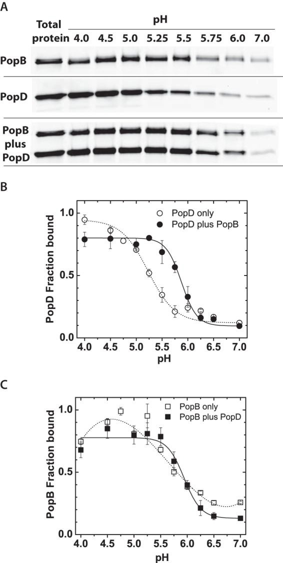
FIGURE 2.

PopB enhanced PopD membrane binding. A, SDS-PAGE gel showing the amount of purified PopDBpyFL and/or PopBBpyFL isolated in the membrane containing fraction after bound and unbound proteins were separated using a liposome flotation membrane-binding assay as described in the text. Total lipids were 2 mm and total protein was 400 nm. Individual proteins or pre-mixed proteins were incubated with membranes at 20–23 °C for 1 h before ultracentrifugation using a sucrose gradient. Proteins were visualized using BpyFL fluorescence. B, PopDBpyFL binding at the indicated pH when the translocator was incubated with membranes alone or when premixed and incubated with an equimolar amount of PopBWT. C, PopBBpyFL binding at the indicated pH when the translocator was incubated with membranes alone or when premixed and incubated with an equimolar amount of PopDWT. BpyFL fluorescence was quantified by gel densitometry and each data point represents the average of at least two independent assays and error bars indicate the data range. Average lines to individual translocators (dashed) and both translocators (solid) are shown only as a guide for the reader.
