FIGURE 5.
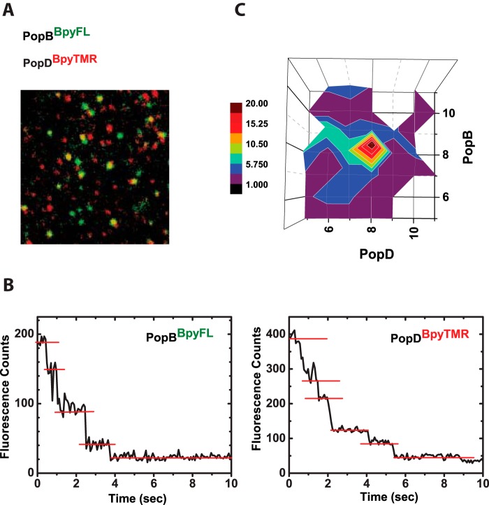
PopB and PopD assemble hexadecameric hetero-complexes in membranes. Protein complexes resulting from the equimolar addition of PopDBpyTMR (red emitting) and PopBBpyFL (green emitting) were imaged using dual color single-molecule TIRF microscopy on SLB. A, merge image of single particles containing PopDBpyTMR (red) and PopBBpyFL (green). Yellow spots indicate co-localization of PopDBpyTMR and PopBBpyFL in individual complexes. B, typical fluorescence intensity time traces for single protein complexes. Photobleaching events appear as stepwise decrease in the intensity. C, quantification of dual-color photobleaching counts obtained from 226 time traces of particles containing both PopDBpyTMR and PopBBpyFL. The heat map for observed PopD:PopB distributions is centered at 8 photobleaching counts for PopD and 8 photobleaching counts for PopB.

