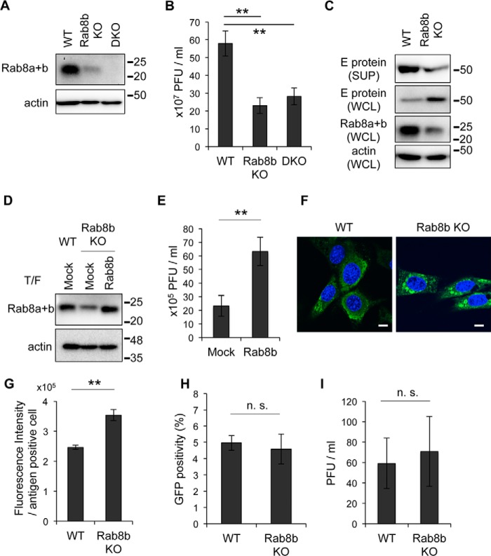
FIGURE 3.
Viral protein is accumulated in cytoplasm in Rab8b KO MEFs. A, Rab8 expression levels in WT, Rab8 KO or DKO MEFs. WT, Rab8b KO, or DKO MEFs were analyzed by immunoblotting for Rab8 and actin. B, viral titer in culture supernatants. WT, Rab8b KO and DKO MEFs were infected with WNV (1 Pfu/cell). Culture supernatants were harvested at 48 hpi, and viral titers were determined by the plaque assay. Data represent mean ± S.D. of three independent experiments. Statistical significance was assessed using Student's t test, and is indicated by asterisks (**, p < 0.01). C, comparison of amount of E protein. Culture supernatants (SUP) and whole cell lysates (WCL) from WNV-infected WT or Rab8b KO MEFs were analyzed by immunoblotting for E protein, Rab8, and actin. These cells were prepared at 48 hpi. D, Rab8a+b expression levels of Rab8b-complemented Rab8b KO MEFs. Control plasmid (Mock) or Rab8b expression plasmid (Rab8b) was introduced into WT or Rab8b KO MEFs, thereafter transfected cells were analyzed by immunoblotting for Rab8 and actin. E, viral titer in culture supernatants from control- or Rab8b expression plasmid-transfected Rab8b KO MEFs. After 24 h, plasmid-transfected cells were infected with WNV (1 Pfu/cell). Culture supernatants were harvested at 48 hpi, and viral titers were measured by plaque assay. Data represent mean ± S.D. of three independent experiments. Statistical significance was assessed using Student's t test, and is indicated by asterisks (**, p < 0.01). F, intracellular localization of E protein. WT or Rab8b KO MEFs were infected with WNV (1 Pfu/cell). Cells were harvested at 48 hpi and stained with E protein (green). Cell nuclei were counterstained with DAPI (blue). Scale bars: 10 μm. G, fluorescence intensity of viral antigen. WNV-infected WT or Rab8b KO MEFs were harvested at 48 hpi and stained with viral antigen. Fluorescence intensity of viral antigen in each cell was analyzed by IN Cell Analyzer. Data represent mean ± S.D. of three independent experiments. Statistical significance was assessed using the Student's t test, and is indicated by asterisks (**, p < 0.01). H, Rab8b does not influence the entry step of WNV infection. WT or Rab8b KO MEFs were inoculated with the VLPs encoding GFP, and GFP positivity was measured by IN Cell Analyzer. Data represent mean ± S.D. of three independent experiments. Statistical significance was assessed using the Student's t test. I, Rab8b does not affect replication of IFV. WT or Rab8b KO MEFs were inoculated with the IFV (1Pfu/cell). The culture supernatants were harvested at 24 hpi, and viral titers were measured by plaque assay. Data represent mean ± S.D. of three independent experiments. Statistical significance was assessed using Student's t test.

