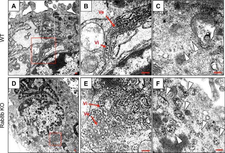
FIGURE 6.
Ultrastructural analysis of WNV-infected WT or Rab8b KO MEFs. Ultrathin sections of WNV-infected, Epon-embedded WT or Rab8b MEFs fixed at 48 hpi are shown in (A–C) or (D–F), respectively. The red and white-boxed areas in A are shown at higher magnification in B and C, respectively. The red and white-boxed areas in D are shown at higher magnification in E and F, respectively. Arrowheads in C and F indicate vesicles containing viral particles. Scale bars: 200 nm.

