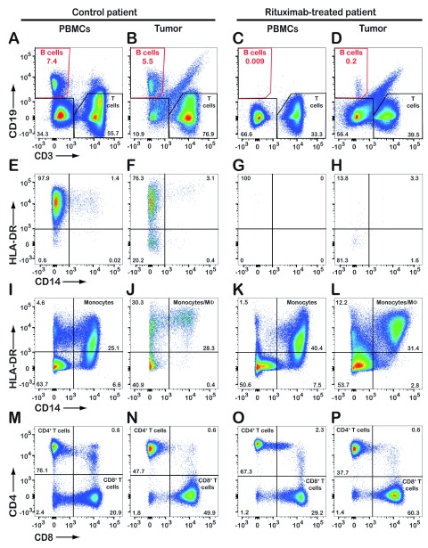
Figure 1. Flow cytometric analysis reveals the absence of tumor-infiltrating CD19 + B cells in a patient treated with rituximab.
Upon chest surgery for removal of a lung tumor from a patient previously treated with rituximab, tumor biopsy and serum samples were analyzed by flow cytometry. Results from a control lung cancer patient (not treated with rituximab) are shown for comparison. Both patients were diagnosed with lung adenocarcinoma. CD45-positive leukocytes were gated and analyzed further for expression of CD19/CD3 ( A– D), HLA-DR/CD14 ( E– L), and CD4/CD8 ( M– P). The dot plots E– H show expression of HLA-DR and CD14 by CD19-positive B cells only (red gates in A– D). Numbers in quadrants indicate the percentage of cells detected. PBMCs, peripheral blood mononuclear cells.

