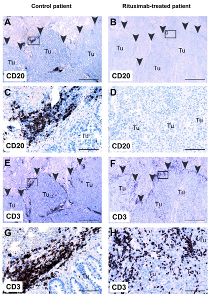
Figure 3. Absence of CD20 + B cells in primary lung tumor from a patient treated with rituximab.
Lung tissue sections from a control patient ( left) and from a rituximab-treated patient ( right) were stained with anti-CD20 ( A– D) or anti-CD3 ( E– H) antibodies, and contrastained with hematoxylin. Both patients were diagnosed with lung adenocarcinoma. Arrowheads delineate the border of the tumor. Small boxes in A, B, E, and F indicate magnified areas in C, D, G, and H, respectively. Tu, tumor tissue. A, B, E, and F: 20× magnification; scalebar = 1 mm. C, D, G, and H: 200× magnification; scalebar = 100 μm.

